Scientists discover brain's immune cells are replaced by more inflammatory blood-derived cells between ages 50 and 75
A major shift in brain immunology challenges the long-held assumption that microglia persist unchanged for life.
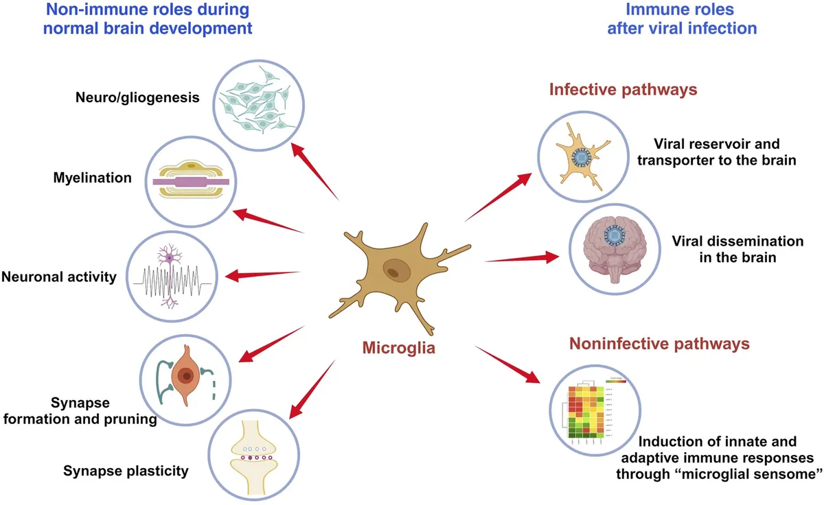
The human brain undergoes a sweeping reorganization of its immune landscape during midlife, with one of the most dramatic shifts occurring between approximately ages 50 and 75. During this window, many of the brain’s original immune cells decline and are replaced by cells with more inflammatory characteristics, according to research published this month. The findings revise a fundamental assumption in neuroscience: that microglia, the brain’s resident immune cells, are stable populations established during embryonic development and maintained unchanged throughout life.
The study identifies a specific population of microglia that originate during embryonic development and decline substantially during this midlife period. These cells are replaced by others with molecular signatures that resemble immune cells from the blood, rather than the brain’s traditional resident immune population. This replacement represents not merely a numerical decline but a qualitative change in the brain’s immunological character.
The inflammatory profile of the incoming cells carries significant implications for understanding brain aging and disease susceptibility. Microglia perform essential maintenance functions—pruning synapses, clearing debris, and responding to injury—but they also shape the brain’s inflammatory environment. A shift toward blood-derived cells with more inflammatory signatures suggests the aging brain may operate under chronically elevated immune activation, even in the absence of specific pathogens or injury.
What makes this finding particularly notable is the precision of the timing. The 50-to-75 age window captures a period when risk for neurodegenerative conditions begins accelerating dramatically, yet the biological mechanisms connecting normal aging to disease vulnerability remain poorly understood. The replacement of embryonic-origin microglia with blood-derived alternatives provides a cellular substrate for that vulnerability—one that operates independently of the protein aggregates or vascular changes typically emphasized in aging research.
The research also raises methodological questions for the field. Studies of brain immunology have historically assumed that microglial populations sampled from older adults represent the same cell type present since early development. If substantial replacement has occurred, comparisons between young and old brains may be conflating fundamentally different cell lineages with distinct functional capacities and regulatory sensitivities. The assumption of cellular continuity across the lifespan, embedded in decades of experimental design, now requires explicit verification.
Whether this immune cell replacement is adaptive, maladaptive, or simply neutral remains to be determined. The inflammatory characteristics of the incoming cells could represent a compensatory response to declining function of original microglia, or they could contribute directly to the neuroinflammatory environments associated with cognitive decline. Either way, the discovery establishes a new biological boundary in human aging—one that sits squarely in midlife and redefines what “normal” brain immunology looks like across the decades.